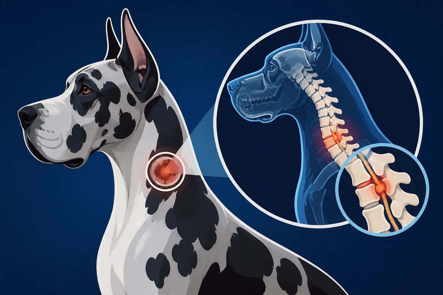
Imaging a Great Dane Neck for Wobblers
Discovering your beloved Great Dane struggling with their gait, exhibiting an unsteady walk, or even collapsing can be utterly heartbreaking and profoundly alarming. It’s a terrifying scenario no owner wants to face, often leaving you scrambling for answers. This comprehensive guide will walk you through the critical imaging processes used to diagnose Wobbler Syndrome in Great Danes, detailing what each scan reveals, why it's indispensable, and what you can expect during this challenging diagnostic journey.
Understanding Wobbler Syndrome in Great Danes
Wobbler Syndrome, a condition that strikes fear into the hearts of many large breed owners, is a complex neurological disorder. Getting to grips with what it actually entails is the first step toward understanding why intricate imaging is so vital.
What is Cervical Spondylomyelopathy?
The official, more scientific name for Wobbler Syndrome is Cervical Spondylomyelopathy (CSM). It’s a mouthful, I know, but it perfectly describes what’s happening. In simple terms, it refers to a condition where the spinal cord in the neck (cervical spine) is compressed. This compression can be caused by a variety of factors, including abnormal bone growth, bulging or herniated discs, or thickened ligaments within the spinal canal. When the spinal cord, which is essentially the highway for nerve signals from the brain to the rest of the body, gets squeezed, those signals can’t get through properly.
For Great Danes, this condition is particularly prevalent due to their rapid growth rate and the unique anatomy of their cervical vertebrae. The vertebral column is made up of individual bones (vertebrae) stacked on top of each other, and in Wobblers, one or more of these vertebrae, or the discs between them, fail to develop or maintain their normal structure, leading to that problematic compression. This is The Neck Condition That Trips a Great Dane Up, making everyday movements difficult and coordination a constant challenge for them.
Recognizing the Early Signs
Catching Wobbler Syndrome early can make a significant difference in management and prognosis. However, the initial signs can be incredibly subtle, often dismissed as clumsy puppy behavior or general stiffness. You might first notice your Great Dane scuffing their back paws when they walk, or their nails might appear unusually worn down on the top. This is a classic indicator that they aren't lifting their feet properly. You might also observe a slightly uncoordinated gait, sometimes described as a "drunken sailor" walk or a distinct wobbly appearance, especially in their hindquarters. This unsteadiness might become more pronounced when they're turning or trying to navigate stairs.
As the condition progresses, these signs typically become more obvious. They might struggle to stand up after lying down, seem reluctant to go for walks, or even start to trip and fall more frequently. Some dogs might develop a shortened stride in their front legs, or carry their head low, signaling neck pain. Recognizing The First Wobbler Signs in a Great Dane and paying attention to subtle clues like Scuffed Back Nails on a Great Dane is crucial. It’s a slow, insidious progression for some, while for others, the decline can be quite rapid. Any persistent change in your Dane's coordination or movement warrants immediate veterinary attention.
Why Imaging is Non-Negotiable
Given the complex nature of Wobbler Syndrome and the various ways it can manifest, a physical examination alone, no matter how thorough, simply isn't enough to secure a definitive diagnosis. Your veterinarian might suspect Wobblers based on clinical signs and neurological tests, but they cannot pinpoint the exact location or cause of the spinal cord compression without advanced imaging. Think of it this way: a physical exam can tell you that there's a problem, but imaging tells you where the problem is and what kind of problem it is.
Precise imaging is absolutely critical because it dictates the entire treatment plan. Without knowing if the compression is caused by a bulging disc, a bony spur, or an unstable vertebra, it's impossible to determine the most effective course of action. Surgical interventions, for instance, are highly specific and require exact knowledge of the anatomy involved. Attempting surgery without this information would be like trying to navigate a complex maze blindfolded. Imaging removes the guesswork, allowing veterinarians and specialists to develop a targeted, effective strategy, whether that’s conservative management or a highly specialized surgical procedure.
The Initial Steps: X-Rays and Their Limitations
When your Great Dane first starts showing those concerning signs, your regular vet will likely begin with routine diagnostic steps, and X-rays are almost always among the first. They offer a quick, accessible look at the skeletal structure, but it’s important to understand what they can and cannot tell us about Wobbler Syndrome.
What Plain Radiographs Can (and Can't) Show
Plain radiographs, more commonly known as X-rays, are a staple in veterinary diagnostics. They're relatively inexpensive, readily available, and non-invasive, meaning your Great Dane usually won't require deep sedation, often just a mild tranquilizer to help them hold still. For a giant breed like a Great Dane, getting them into the right position for a good X-ray can be a bit of a dance, but it's manageable.
What X-rays can show us are the bones. They're excellent for visualizing gross bone abnormalities, such as obvious fractures, severe arthritis, or significant malformations of the vertebral bodies themselves. In the context of Wobbler Syndrome, they might reveal some bony changes that suggest instability or narrowing of the spinal canal. For example, they can show if vertebrae are misaligned or if there's evidence of bone spurs that could be encroaching on the spinal cord.
However, and this is a big "however," X-rays are notoriously poor at visualizing soft tissues. The spinal cord itself, along with the discs between the vertebrae and the ligaments that support the spine, simply don't show up clearly on a standard X-ray. This means that while X-rays might give us clues about potential bony causes of compression, they cannot definitively confirm that the spinal cord is compressed, nor can they show the severity of that compression. It's like looking at the outside of a house and trying to guess what the plumbing looks like inside – you can infer some things, but you don't have the full picture.
Dynamic X-Rays: A Glimpse into Movement
To try and overcome some of the limitations of plain X-rays, veterinarians might suggest dynamic X-rays. These involve taking X-ray images while your Great Dane’s neck is positioned in different ways – typically in full flexion (head tucked down) and full extension (head stretched up). The idea behind this is to assess the stability of the cervical spine. In some cases of Wobbler Syndrome, the spinal cord compression only becomes apparent or worsens when the neck is moved into certain positions. This instability is a critical factor, as it means the spinal cord is being squished and released with every movement, leading to chronic irritation and damage.
Performing dynamic X-rays on a large breed like a Great Dane requires careful handling and usually a more significant level of sedation or even light anesthesia to ensure the dog remains perfectly still in precise, and sometimes uncomfortable, positions. The veterinary team must be very experienced to minimize stress and ensure the safety of your Dane, especially considering that manipulating an already compromised neck carries a small risk. While dynamic views can sometimes highlight areas of instability that might be missed on static X-rays, they still suffer from the same fundamental limitation: they cannot directly visualize the soft tissues of the spinal cord or precisely quantify the degree of compression. They're a step up, but not the final answer.
Why More Advanced Imaging is Often Needed
Ultimately, while X-rays provide a good starting point and can rule out other bone-related issues, they rarely provide sufficient detail for a definitive diagnosis of Wobbler Syndrome or, more importantly, for precise surgical planning. Think of them as the initial screening tool. They can tell you if there’s smoke, but not the exact source of the fire or how big it is.
For Great Danes presenting with neurological signs suggestive of Wobblers, it’s almost always necessary to move to more advanced imaging techniques. These sophisticated scans offer a cross-sectional view of the neck, allowing veterinary specialists to look at the spinal cord, discs, and surrounding soft tissues in incredible detail. Without this level of detail, choosing the correct treatment – especially if surgery is on the table – becomes a dangerous guessing game. This means that after initial X-rays, your vet will likely recommend a referral to a veterinary neurologist for further, more specialized diagnostics.
The Gold Standard: MRI and CT Scans
When it comes to truly understanding what's going on inside your Great Dane's neck and accurately diagnosing Wobbler Syndrome, Magnetic Resonance Imaging (MRI) and Computed Tomography (CT) scans are the gold standard. These advanced imaging techniques provide the detailed cross-sectional views that traditional X-rays simply can't.
Magnetic Resonance Imaging (MRI): The Soft Tissue Specialist
If Wobbler Syndrome is suspected, an MRI is often the preferred diagnostic tool. MRI technology is absolutely incredible because it uses powerful magnetic fields and radio waves, rather than X-rays, to create exquisitely detailed images of soft tissues. This means that for conditions like Wobblers, an MRI can clearly visualize the spinal cord itself, the intervertebral discs, ligaments, and nerves – all the structures that are typically affected by compression.
An MRI will show the exact location of any spinal cord compression, the severity of that compression, and precisely what is causing it. Is it a herniated disc bulging into the spinal canal? Is it a thickened ligament? Or is it a bony abnormality pushing on the cord? The MRI will reveal these details, which are absolutely paramount for guiding treatment decisions. Without this clarity, effective surgical planning would be impossible. You'll literally see What Shows Up on a Great Dane Spinal Scan, giving you and your veterinary team a clear picture of the internal structures.
The downside? An MRI requires your Great Dane to be completely still for an extended period, which means general anesthesia is a must. This naturally brings up concerns about anesthesia risks, especially for a giant breed. We'll delve more into that soon, but it's an important consideration. Also, MRI scans are costly. We're talking several thousand dollars, depending on your location and the specific veterinary specialty hospital. This is where having robust pet insurance, like Healthy Paws, can be a true lifesaver, helping to cover a significant portion of these substantial diagnostic costs.
Computed Tomography (CT) Scan: Bone Detail and Surgical Planning
While MRI excels at soft tissue visualization, a CT scan takes the crown when it comes to detailing bony structures. A CT scanner uses a series of X-ray beams taken from multiple angles, which a computer then processes to create detailed cross-sectional images, almost like slices of bread from a loaf. It's particularly adept at showing bone spurs, malformed vertebrae, and subtle joint changes that might be contributing to spinal cord compression.
In the context of Wobbler Syndrome, a CT scan is invaluable for surgical planning. Surgeons often use CT images to get a precise, three-dimensional understanding of the bony architecture around the compressed spinal cord. This allows them to visualize the exact angles and approaches needed for surgical intervention, potentially minimizing risks and improving outcomes. Sometimes, both an MRI and a CT scan are performed, as they complement each other perfectly: the MRI provides the crucial soft tissue detail, while the CT offers the bony roadmap for surgery.
Like MRI, a CT scan also requires general anesthesia for your Great Dane. While generally quicker than an MRI, it still demands a motionless patient. The cost is also substantial, though often slightly less than an MRI alone. Many owners find that their pet insurance, such as Healthy Paws, is an invaluable tool in managing the financial burden associated with these high-tech diagnostic procedures, helping ensure their beloved Dane gets the best possible care without undue financial strain.
Myelography: An Older, Riskier Technique
Before the widespread availability and sophistication of MRI and CT scans, myelography was the primary advanced imaging technique used to diagnose spinal cord compression. It involves injecting a special contrast dye directly into the space surrounding the spinal cord (the subarachnoid space). Once injected, the dye flows around the spinal cord, and X-rays or CT scans are then taken. Areas where the spinal cord is compressed will show up as an interruption or narrowing of the dye column, essentially outlining the compression.
While myelography can still be performed and is sometimes used if MRI or CT is unavailable or cost-prohibitive, it carries significantly higher risks compared to non-invasive MRI or CT. The injection of dye into the spinal canal can cause adverse reactions, including seizures, particularly in Great Danes, who can be more susceptible to neurological sensitivities. It can also, in rare cases, worsen existing neurological signs or introduce new ones. Because of these risks and the superior detail provided by MRI (especially for soft tissues), myelography has largely been superseded by MRI and CT as the preferred diagnostic method for Wobbler Syndrome. Most veterinary neurologists will opt for MRI or CT whenever possible to ensure both the safety of the patient and the accuracy of the diagnosis.
Preparing Your Great Dane for Advanced Imaging
Deciding to go forward with advanced imaging for your Great Dane is a big step. It’s not just about the cost; it’s about the logistics, the emotional toll, and the concerns about your giant companion undergoing anesthesia. Being prepared for these aspects can help ease the process.
The Anesthesia Conversation
One of the biggest concerns for any Great Dane owner facing advanced imaging is the need for general anesthesia. Great Danes, like many large and giant breeds, can be more susceptible to certain anesthetic risks, and their sheer size means higher drug dosages and a greater physiological impact. This is not to scare you, but to highlight why it’s absolutely critical to have a thorough discussion with your veterinary team about the anesthesia protocol.
Before any procedure requiring anesthesia, your Great Dane will undergo comprehensive pre-anesthetic bloodwork to assess organ function (kidneys, liver) and ensure they are healthy enough for the procedure. An electrocardiogram (ECG) might also be recommended to check heart function, especially given that Great Danes are prone to certain cardiac conditions like dilated cardiomyopathy. The veterinary team will typically place an intravenous (IV) catheter to administer fluids during the procedure, which helps maintain blood pressure and supports organ function.
The good news is that veterinary anesthesia has advanced dramatically. Modern anesthetic agents are safer, and monitoring equipment is incredibly sophisticated. Your Great Dane will be continuously monitored by a dedicated veterinary technician, checking vital signs like heart rate, breathing rate, oxygen saturation, blood pressure, and temperature throughout the entire procedure. Don’t hesitate to ask about the experience level of the anesthetist, the monitoring equipment used, and any specific protocols they have for giant breeds. Understanding the process can significantly alleviate your anxiety regarding Anesthesia Risk in a Great Dane. Remember, covering these diagnostic and anesthetic costs is precisely what pet insurance providers like Healthy Paws are designed to help with, making these vital procedures more accessible.
Logistics and Costs
Let's not sugarcoat it: advanced imaging for a Great Dane is a significant financial undertaking. As mentioned, an MRI or CT scan can easily run into the thousands of dollars, a figure that can feel overwhelming when your heart is already heavy with worry for your dog. This cost typically includes the anesthesia, the scan itself, and the interpretation by a board-certified veterinary radiologist or neurologist. It's often one of the largest single expenses a Great Dane owner will face for a health issue. To help understand the full financial scope, you might find it helpful to look into articles like Paying for a Great Dane Wobbler Workup.
Beyond the direct costs, there are logistical considerations. Most advanced imaging for Great Danes with suspected Wobbler Syndrome will happen at a specialty veterinary hospital, which might be a significant drive from your home. You'll need to plan for travel, drop-off, and pick-up. Sometimes, depending on the schedule and your Dane's recovery from anesthesia, an overnight stay might be recommended. Ensure you discuss the full scope of costs, including any potential overnight fees, with the specialty clinic beforehand.
This is precisely where having pet insurance, such as Healthy Paws, proves to be invaluable. Many owners opt for comprehensive plans specifically to cover unexpected illnesses and accidents, which includes advanced diagnostics like MRI and CT scans. Being able to submit a claim for a substantial portion of these bills can provide immense relief during an already stressful time, allowing you to focus on your Great Dane’s health rather than the crushing weight of the veterinary bill.
What Happens During and After the Scan
On the day of the scan, your Great Dane will be admitted to the specialty hospital. They'll typically be fasted (no food or water) for several hours prior to the anesthesia, as instructed by your vet. Once admitted, the veterinary team will perform their pre-anesthetic checks, and then your Dane will be gently sedated and intubated to ensure a clear airway.
During the scan, whether it's an MRI or CT, your Great Dane will be carefully positioned on a special table that slides into the imaging machine. The team will ensure precise alignment to capture the best possible images. Advanced monitoring equipment will continuously track their vital signs. The actual scanning process can take anywhere from 30 minutes to over an hour, depending on the type of scan and the number of images required. While the machines can be quite loud, your Dane will be comfortably asleep and unaware.
After the scan is complete, your Great Dane will be moved to a recovery area where the veterinary team will closely monitor them as they wake up from anesthesia. They might be a bit groggy, disoriented, or wobbly (ironically!) as the anesthetic drugs wear off. This recovery period is critical for ensuring they wake up safely and without complications. You'll typically receive a call once they are fully recovered and ready for discharge. The images will then be reviewed by a specialist, and your referring veterinarian or the neurologist will contact you to discuss the findings and the proposed next steps. Patience is key during this waiting period – it’s often the hardest part.
Interpreting the Results and Next Steps
Once the advanced imaging is complete, the images are meticulously reviewed, usually by a board-certified veterinary radiologist or neurologist. This interpretation is where all the puzzle pieces finally start to come together, leading to a clear diagnosis and a concrete plan for your Great Dane.
Understanding the Diagnosis
The specialist will carefully analyze every "slice" of the MRI or CT scan, looking for any abnormalities that could be compressing the spinal cord. They are searching for specific patterns of compression, such as disc herniations (where the soft cushion between vertebrae bulges or ruptures), bony malformations (abnormal growth of the vertebrae themselves), or hypertrophy of ligaments (thickening of the connective tissues that support the spine). For Great Danes, it's common to see a combination of these factors contributing to the problem. The precise anatomy of Why Great Dane Neck Vertebrae Pinch the Cord is unique, and these advanced scans pinpoint the exact location and nature of this pinch.
They'll identify not only what is causing the compression but also where it is occurring (e.g., between C5 and C6 vertebrae) and how severely the spinal cord is affected. This detailed information is crucial because Wobbler Syndrome isn't a single condition but rather a spectrum of issues all leading to the same clinical signs. Understanding these nuances is essential for tailoring the most effective treatment. The diagnosis will give you a clear picture of what your Great Dane is facing.
Weighing Treatment Options
With a definitive diagnosis in hand, your veterinary neurologist will discuss the various treatment options available for your Great Dane. These generally fall into two broad categories: conservative medical management or surgical intervention.
Conservative management typically involves a combination of anti-inflammatory medications, pain relief, activity restriction (often crate rest), and sometimes physical therapy. This approach is usually considered for Great Danes with mild signs, or those who are not candidates for surgery due to other health issues or advanced age.
However, for many Great Danes with Wobbler Syndrome, especially those with moderate to severe neurological deficits or progressive signs, surgery is often recommended as the most effective long-term solution. There are several different surgical techniques, each designed to decompress the spinal cord by addressing the specific cause (e.g., removing a herniated disc, stabilizing unstable vertebrae, or excising bony spurs). The choice of surgical procedure is entirely dependent on the detailed information provided by the MRI or CT scan. Without this precise imaging, a surgeon would not know which approach to take or even if surgery would be beneficial. Articles discussing Surgery Against Management for a Wobbler Dane highlight this critical decision point.
Prognosis and Ongoing Management
The prognosis for a Great Dane with Wobbler Syndrome varies widely depending on several factors: the severity of the spinal cord compression, the type of compression, the duration of clinical signs before diagnosis, and the chosen treatment method. For dogs undergoing successful surgery, there's often a good chance of improvement in neurological function, though full recovery isn't always guaranteed, and some residual wobbliness may remain. For those managed conservatively, the goal is often to stabilize the condition and maintain comfort, rather than achieve significant improvement.
Wobbler Syndrome is rarely a "one and done" situation. It's often a lifelong condition that requires ongoing management, regardless of whether surgery is performed. This can include long-term medication, regular physical therapy sessions, lifestyle modifications (e.g., using a harness instead of a collar, avoiding jumping or strenuous activity), and routine follow-up appointments with your veterinarian or neurologist. It's a journey that demands dedication and patience from both you and your Great Dane, but with accurate diagnosis through imaging and appropriate treatment, many Great Danes can still enjoy a good quality of life.
Comparison of Imaging Techniques for Wobbler Syndrome
To help you visualize the differences, here's a table summarizing the main characteristics of the imaging techniques discussed for diagnosing Wobbler Syndrome in Great Danes:
| Imaging Technique | Primary Use for Wobblers | Detail Level | Anesthesia Required | Cost Range (Relative) | Key Advantages | Key Disadvantages |
|---|---|---|---|---|---|---|
| Plain X-rays | Initial screening, bone abnormalities | Low | Mild Sedation (often) | Low | Quick, widely available, inexpensive | Poor soft tissue detail, cannot confirm spinal cord compression |
| Dynamic X-rays | Assess spinal instability | Low to Moderate | Moderate Sedation/Anesthesia | Low to Moderate | Can show instability with movement | Still poor soft tissue detail, risks with manipulation |
| CT Scan | Bony compression, surgical planning | High (bone) | General Anesthesia | Moderate to High | Excellent bone detail, 3D reconstruction, quicker than MRI | Limited soft tissue detail compared to MRI, uses radiation |
| MRI Scan | Soft tissue compression, definitive diagnosis | High (soft tissue) | General Anesthesia | High | Superior soft tissue detail (spinal cord, discs), no radiation | Longer procedure, very expensive, requires specialized equipment |
| Myelography | Spinal cord compression (older method) | Moderate | General Anesthesia | Moderate | Can outline compression when MRI/CT unavailable | Invasive, potential for neurological side effects (seizures), uses radiation |
Please note that costs are relative and can vary significantly based on location, facility, and the specific needs of your Great Dane. Always consult with your veterinarian for precise estimates.
Disclaimer
The information provided in this guide is intended for educational purposes only and should not be considered a substitute for professional veterinary advice. Always consult with a qualified veterinarian or veterinary specialist for any health concerns or before making any decisions about your Great Dane's medical care.